

INFRARED IMAGING SYSTEMS AND METHODS
Our team has 30 years of experience in the development of infrared thermal imaging systems and technologies. Since 1992, 12 original models of thermal imagers have been developed, and more than 30 devices have been prototyped for solving various tasks of thermal analysis. We also have significant experience in developing thermal imaging techniques and applying the thermal imaging method in diverse fields of human activity, including science and medicine. The main directions of our scientific activity today are:
Development of infrared image converters based on superconductors
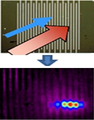 We study bolometric, noise and other properties of various superconducting film structures, as well as the mechanisms of formation of their response to electromagnetic radiation to create effective infrared image converters in a wide spectral range. The principle of operation of the converters is based on the original idea of controlling the coordinate sensitivity of a superconducting bolometric structure using an external (current, magnetic, thermal, etc.) influence. The method is experimentally implemented using a laser scanning beam, which forms areas sensitive to IR radiation on the surface of a HTSC thin-film meander. A HTSC bolometric multi-element IR image converter with electrodeless readout of the thermal field distribution has been created, which is an alternative to IR thermal imaging systems based on multi-element matrices. [1, 2, 3]
Creation of original models of IR systems
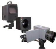 We create various models of IR thermal imaging systems that are capable of simultaneously solving both standard thermal imaging analysis tasks and diverse atypical tasks that arise in scientific research (for example, high or low-temperature processes). Such tasks cannot be solved by serial thermal imagers because they are not available for changes and modernization. A distinctive feature of our models is the "open architecture" and modular design of the hardware and software parts, which allows you to change parameters and functionality in accordance with the specifics of the task, combine the device with other scientific or medical equipment, etc. [4, 5, 6, 7]
Study of the thermal field dynamics
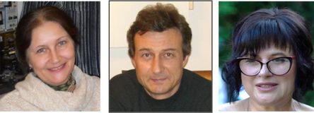
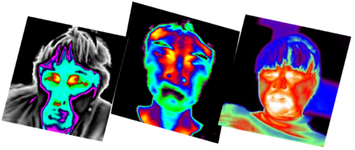
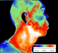 We study the distribution and dynamics of abnormal thermal fields on the skin surface of cancer patients for early non-invasive diagnosis of malignant tumors, as well as for the prediction and current control of the individual level of side toxic reactions that occur during radiation therapy. [8, 9]
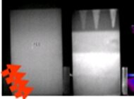 We use the dynamic thermography method to measure the amplitude and temporal changes of excess temperature fields on the surface of aircraft parts and other products made of composite materials in order to detect internal defects and quantify their parameters (defect shape, size, depth of occurrence and material). [10]
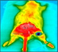 We study the dynamics of temperature fields on the surface of model systems and biological objects during cryoexposure of various regimes (durations, temperature, etc.) in order to develop a thermal imaging procedure of intraoperative monitoring of the growth of necrosis zone in pathological tissue. [11, 12]
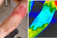 We study thermal fields on the skin surface of patients with soft tissue injuries (wounds, burns, frostbite, soft tissue infection) for the purpose of early diagnosis of the depth of the lesion and monitoring the treatment process, identifying areas of necrosis, circulatory disorders, inflammation and other pathological processes. [13]